
Laser Therapy in Diabetic Macular Edema
Keshav B.R., George Zacharia, Varsha K. Bhat, Mary K. Joseph, Thara Ideculla
ABSTRACT
Introduction: Diabetic macular edema results in irreversible loss of vision and is the major cause of visual morbidity in patients with Diabetes of adult onset. DCCT trial has linked this to poorer control and increased duration of DM. laser treatment in such cases is known to reduce visual impairment by 50% for a period of 5 years. 3 The aim of this study is to evaluate the effect of laser on visual outcome in patients with clinically significant macular edema and also evaluate the effect of some factors like control of blood sugar, hypertension, nephropathy etc.
Methods: Retrospective analysis of 165 eyes of patients with diabetic maculopathy who underwent laser as per the ETDRS (early treatment diabetic retinopathy study) protocol 1 was made. All these patients underwent Visual Acuity check, slit lamp examination of anterior segment, IOP check and after dilatation detailed examination of macula with 78/90 d lens and areas of retinal thickening recorded and subjected to focal laser/grid laser depending upon whether the maculopathy was focal/diffuse. Patients were assessed for control of sugar and presence of hypertension/nephropathy or any other related systemic diseases. Patients were followed up for a minimum of 6 months to a maximum of 24 months. The visual acuity at the end of 3-4 months was taken as final visual acuity after laser.
Results: 165 eyes of patients having CSME (clinically significant macular edema) were subjected to laser. 108 (64.54%) eyes underwent focal laser and the rest were given grid laser. 153 eyes underwent macular laser without PRP (Pan retinal photocoagulation) while rest had even PRP along with macular laser. 92 eyes (55.75%) totally, 39 eyes (59.1%) in controlled group and 26 eyes (56.5%) in the controlled group and 12 eyes (54.5%) in patients with hypertension had stable vision 3-4 months after laser. 44 eyes (26.7%) overall, 18 eyes (27.3%) in controlled group, 16 eyes (34.8%) in uncontrolled group and 7 eyes (31.8%) in hypertensive showed improvement of vision after laser.
Conclusion: More than 50% of eyes of patients who underwent laser had stabilization of VA and >25% of eyes had improvement in VA. No correlation was found between outcome of laser and control of blood sugar and associated hypertension.
Keywords: CSME, Focal laser, Grid laser, ETDRS (early treatment diabetic retinopathy study)
Submitted: 14 June 2007
Reviewed: 2 August 2007
Accepted: 25 September 2007
From the Department of Ophthalmology, Sur Regional Hospital
Address Correspondence and reprint request to: : Dr. BR Keshav, H.O.D Ophthalmology, Sur Hospital
E-mail:keshavraj_2000@yahoo.com
INTRODUCTION
Diabetic macular edema is one of the common causes of visual impairment in diabetes. Overall macular edema accounts for more vision impairment than PDR (proliferative diabetic retinopathy) in people with diabetes. 4 Laser treatment for clinically significant macular edema has been the accepted form of treatment worldwide after the recommendations by the ETDRS study. 1 Laser treatment is known to reduce further visual impairment in patients with diabetic macular edema for a period of 5 years1 as compared to those who had no such treatment making it an important modality of treatment. In this study an attempt has been made to analyse the results of lasers in cases of clinically significant macular edema attending our clinic in Sur hospital and also try to compare the results with respect to control of diabetes, with/without renal complications, presence of hypertension or not and so on so as to arrive at some sort of conclusion that may help us in counseling our patients and tell them as to the sort of outcome that they may expect after laser therapy.The aim of the study is to analyse the visual outcome in patients with diabetic macular edema after laser therapy, and to compare the visual outcome in these patients with respect to control of diabetes, duration of DM, nephropathy and hypertension
METHODS
One hundred and sixty five eyes of patients attending the eye clinic of Sur hospital from October 2000 to Nov 2006 were included in the study. In each of these patients; history as regards to duration of DM, presence of hypertension/nephropathy was taken. Other relevant history like d/v, treatment that was being taken for DM and so on were taken and recorded. All these patients were subjected to VA check and a detailed anterior segment examination by slit lamp, IOP recorded and detailed fundus examination with indirect ophthalmoscope and 78D lens after full dilatation was made. In each of these cases areas of thickening was noted and classified as diffuse or focal areas of thickening and those cases falling into the purview of clinically significant macular edema as defined by the ETDRS were scheduled for laser. Cases with focal edema were subjected to focal laser and cases with diffuse edema were subjected to macular grid laser. 1
An improvement by one or more lines on Snellens “E” chart on more than one visit, 2 months after laser was taken as significant. Control of DM was considered if the FBS was normal 2 weeks before and 2 weeks after the laser therapy.
RESULTS
One hundred and sixty five eyes of patients attending the eye clinic of Sur hospital with a diagnosis of clinically significant macular edema were subjected to either focal or grid laser and followed up for evaluating the outcome and the results were compiled. One hundred and one (61.25%) were males and 64 (38.8%) were females. (Table 1)
Table 1: Shows age and sex distribution of the cases
Age in yrs |
Male |
Female |
Total |
<40 |
2 |
1 |
3(1.8%) |
40-60 |
55 |
47 |
102(61.8%) |
>60 |
44 |
16 |
50(30.3%) |
Total |
101(61.2%) |
64 (38.8%) |
|
One hundred and two patients (61.8%) were between 40-60 years and 60 patients (36.36%) were above 60 years of age. A negligible number (3 patients) were <40 years. (Table 1)
46 patients (28%) were having normal blood sugar levels, 66 patients (40%) were uncontrolled and in 53 patients (32%) the exact diabetic control was not made out. (Table 2)
Table 2: Shows proportion of uncontrolled cases and controlled cases
Controlled |
Uncontrolled |
Not known |
46 |
66 |
53 |
27.9% |
40% |
32.1% |
Fourty two patients (25.45%) had diabetes for 5-10 years, 72 patients (43.63%) had a duration of 10-20 years, 10 patients (6%) had DM for >20 years and a small number of patients (13-7.8%) had DM of <5 years. In around 28 patients (16.96%) the duration was not known.(Table 3)
Table 3: Patients with other systemic diseases
Hypertension |
Nephropathy |
CCF |
22 |
7 |
3 |
13.33% |
4.24% |
1.8% |
CCF: Congestive Cardiac Failure |
||
As regards to systemic problems other than DM in these patients a very few of them had hypertension 22 patients (13%), nephropathy 7 patients (4.24%) and CCF 3 patients (1.8%). (Table 4)
Table 4: Shows the proportion of patients with different types of laser treatment
Focal laser |
Grid laser |
108 |
57 |
65.45% |
34.55% |
One hundred and eight eyes of patients (65.45%) underwent focal laser and 57 eyes of patients (34.55%) underwent grid laser. (Table 5)
Table 5: Shows patients who underwent Pan retinal photocoagulation along with macular treatment
With PRP |
Without PRP |
12 |
153 |
7.28% |
92.72% |
PRP: Pan Retinal Photocoagulation |
|
153 eyes (92.72%) had only treatment for macular edema while a small proportion of patients i.e., 12 eyes (7.28%) also underwent Pan retinal photocoagulation (PRP). (Table 6)
Table 6: Distribution by duration of Diabetes Mellitus
<5yrs |
5-10yrs |
10-20yrs |
>20yrs |
Not known |
13 |
42 |
72 |
10 |
28 |
7.88% |
25.45% |
43.63% |
6.06% |
16.96% |
When the outcome of lasers were analyzed 92 eyes (55.75%) had a stable outcome where VA neither increased nor decreased over 3-4 months after laser. 44 eyes (26.7%) showed an improvement of VA by one or more lines on the E chart while 24 eyes (14.54%) showed worsening of VA by one or more lines. Around 5 patients (3%) were lost for follow up. (Graph-1) Thus 82% of eyes in the study had a stable or improvement of VA following laser.
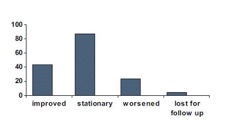
Graph 1: Distribution of patients as per the outcome
When the visual acuity status were analyzed among the patients before and after laser interestingly equal number of patients were seen in various grades of VA namely 53 eyes (32.1%) in the 6/6-6/18 category, 85 eyes (51.5%) in the 6/24-6/60 category and 27 eyes before laser in <6/60 category and 24 eyes in the same category after laser while 3 were lost for follow up. (Graph-2) This suggests that there was no significant shift from one grade of VA to another after laser.
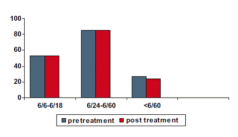
Graph 2: Shows distribution of patients with regards to VA pre and post laser treatment
When the VA status was compared with control of DM, it was found that in the controlled group, majority, 39 eyes (59.1%) had stable outcome while 18 eyes (27.3%) of patients showed improvement in VA and 9 patients 13.6% of eyes of patients showed worsening of VA. In the uncontrolled group also majority of the patients namely 26 eyes (56.5%) had a stable outcome and 16 eyes (34.8%) showed improvement and rest showed worsening. Among the patients with associated Hypertension 12 eyes (54.5%) had stable VA while 7 eyes (31.8%) had improvement in VA and 3 eyes (13.6%) had worsening of VA. (Graph 3) Thus more than half the patients in the controlled group, uncontrolled group and in patients with associated hypertension had a stable outcome in so far as VA is concerned.
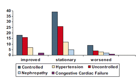
Graph 3: Shows distribution of outcome based on influencing factors
DISCUSSION
Diabetic macular edema is an important cause of visual impairment in diabetics esp. NIDDM and even today laser is one of the most important and most common modality of treatment all over the world despite newer treatments that have emerged in the recent past.
The follow-up of these patients were anywhere from 6 months to 18 months but to assess visual outcome in our cases we restricted our follow-up to 3-4 months as other causes like development of cataract, uncontrolled blood sugar, development of proliferative changes or new macular changes may obscure the outcome of the laser. Although in some studies the follow-up of patients ranged from 6-45 months.2
Regarding the visual outcome it was seen that visual acuity was stabilized at the pre laser level in 55.75% and improved in 27.2%. In a study by Romanuik W. et al.2 it was seen that the stabilization occurred in about similar magnitude i.e. 51.3% and improvement in only 10% as against 27% in our study. The latter probably could be explained due to longer follow-up period in their study. In a study by Lange et al,3 it was shown that stabilization of visual acuity occurred in 50% of cases after laser therapy in 5 years. deterioration of VA was seen in 15% of cases in our study while in Romanuik2 study it was as high as 38.6% which probably can be explained by the difference in the length of follow-up in these studies.
Here an attempt was made to analyse visual outcome in cases where blood sugar was not controlled and where blood sugar was controlled. No such correlation has been made in earlier studies. Level of blood sugar may have a say on hemodynamics and thus retinal capillary permeability so we tried to find a correlation between the two. The conclusion was that 59.1% in the controlled group and 56.5% in the uncontrolled group had stabilization of VA post laser suggesting that there was no significant difference and thus no correlation between outcome of laser treatment and control of blood sugar. In our study Blood sugar levels approximately within 2 wks before or after the laser, where available was considered to come to a conclusion about control of blood sugar while ideally glycosalated Hb would have been a better indicator. A prospective study in this direction will give us better information in this regard.
We also tried to find a correlation between laser outcome and presence of hypertension but it was found that >50% of these cases had stabilization of VA post laser suggesting that there is no correlation. Patients with nephropathy and cardiac problems were in negligible numbers to come to any sort of conclusion.
SUMMARY
A total of 165 eyes were subjected to laser for CSME as per the standard guidelines laid by the ETDRS. 101(61.25%) were males and 64(38.8%) were females. In around 80% of cases the duration of DM ranged from 5-20 years. 92.72% of cases underwent only focal laser while the rest had grid laser. Majority of them had only macular laser without PRP (pan retinal photocoagulation), 22 cases had h/o hypertension, 82% of cases had a stable (55.75%) or improvement (26.7%) in VA (visual acuity), other systemic associations like nephropathy were found in negligible numbers. More than half of the eyes in controlled group, uncontrolled group and patients with associated hypertension had stabilization of VA following laser thus showing that these factors namely control of Blood sugar and hypertension had no influence on the outcome of laser therapy.
CONCLUSION
- More than 50% had stabilization of visual acuity and more than 25% had improvement in visual acuity after laser for CSME
- There was no correlation between visual outcome following laser for CSME and control of blood sugar.
- There was no correlation between outcome of laser and hypertension
REFERENCES
-
No authors listed. “Photocoagulation for diabetic macular edema”. early treatment diabetic retinopathy study report number 1. early treatment diabetic retinopathy research group: Arch ophthalmol, 1985; 103:1796-806.
-
Romaniuk W, Koziol H, Markowska J, Fronczek M. “A grid pattern type of photocoagulation in treatment of diabetic maculopathy-personal experience” Klin Ocanza. 2000; 102:183-6.
-
Lange GE. “laser treatment in diabetic retinopathy”: diabetic retinopathy.Dev ophthalmol 2007; 39:48-68.
-
Klien R, Klien BE, Moss SE et al. The Wisconsin epidemiologic study of diabetic retinopathy. IV. Diabetic macular edema. Ophthalmology.1984; 91:1464-1474.